How One Mark Finds Its Reader
Read the transcript of “How One Mark Finds Its Reader”
Film transcript: How One Mark Finds Its Reader
A receptor writes phosphotyrosine marks across its tail. How can one protein find the right mark among all the others? Phosphate changes a small surface patch, but the mark usually does little alone. It becomes useful when a reader binds there. The reader brings its host protein into the growing signaling complex. A kinase writes the mark, and a phosphatase can erase it. An SH2 domain is one such reader. Its first binding pocket fits phosphotyrosine. Positively charged side chains help grip the phosphate. Unmodified tyrosine lacks that phosphate, so the pocket cannot make the same contact. That first pocket answers whether tyrosine is phosphorylated.
It cannot identify which phosphorylated site this is. A neighbouring surface reads residues beside the phosphotyrosine and supplies another layer of preference. Different SH2 domains carry differently shaped neighbouring surfaces. Now the reader tests both features together. Several tails offer the same gold phosphate. Their neighbouring patterns differ. Matching blocks settle into the groove; poor fits make the reader release.
Src-family readers favour one pattern, while other readers favour others. Here is the rule. Reusable binding domains turn recognized motifs or lipids into protein connections. For SH2, phosphorylation enables the contact, while sequence context helps choose among possible partners. The domain binds; it does not catalyse a reaction. Predict before the reader moves. Both tails carry phosphotyrosine. Only the lower neighbouring pattern fits this reader's groove. Which tail will retain the reader longer? Watch both parts of the binding surface.
The lower tail makes both contacts, so it retains this reader more strongly in this comparison. The upper tail still has phosphate. Its neighbouring pattern simply provides a poorer fit for this domain. Another SH2 domain could prefer a different pattern. The domain is modular because it keeps this binding activity when moved. Detach the reader from one host. Attach it to a different host, and its preferred target remains the same. The new host can bring a different cellular activity to that site. Preference is not destiny. Location, abundance, timing, and the rest of the host protein also shape contacts in cells. The domain narrows the candidates; cellular context narrows them further.
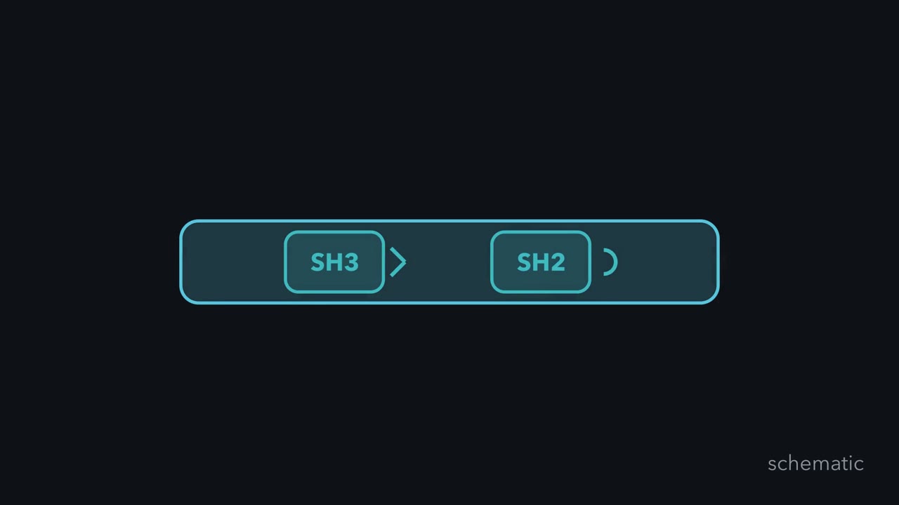
A preferred motif cannot recruit a reader that never reaches it. Other modules apply the same logic to other targets. SH3 reads proline-rich motifs, while some PH domains read membrane lipids. The target changes, but recognition still creates a connection. For PH, that connection is to the membrane.
A receptor has just phosphorylated its own tyrosines. What reads the marks, and how does each reader pick its mark?
Continue in the lesson: Protein Modules: SH2, SH3, PTB, PDZ and PH →
3:41 · Free, with captions and a transcript. The lesson has the full text, the questions and the sources.
More films from this lesson
How this film was made. Narration: written from this lesson's text and read by an AI narrator voice (Kokoro, af_heart). Animation: AI-generated in code (Manim) by Claude, a schematic with no drawn molecular structures. Captions come from the narration script. See something wrong? Report it.