How Conventional PKC Reads Both Branches
Read the transcript of “How Conventional PKC Reads Both Branches”
Film transcript: How Conventional PKC Reads Both Branches
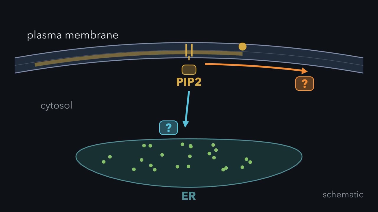
The first film left IP3 traveling and DAG in the membrane. How can conventional protein kinase C use both branches? The branches reach the same cell but keep different locations. Return to the same cell and follow one kinase. Follow its movement and timing.
At rest, conventional PKC is folded shut. Its pseudosubstrate occupies the kinase active site, blocking real substrates. All four parts are connected in one protein chain. A C2 domain can respond to calcium. A C1 domain can recognize DAG. IP3 opened the endoplasmic reticulum channel, raising cytosolic calcium. Calcium binds C2 and changes its membrane affinity. C2 then engages anionic lipids, bringing conventional PKC toward the plasma membrane. The kinase is now close enough to sample the local membrane. At the membrane, C1 can search for DAG. DAG binding strengthens membrane engagement and helps displace the pseudosubstrate. The active site opens, so the kinase can phosphorylate protein substrates.
Now compare three schematic conditions: calcium without DAG, DAG without raised calcium, and both branches together. Which condition should retain conventional PKC most strongly at this membrane? Each contact alone can be weak under physiological conditions. Together, their binding energies add, so membrane preference can rise sharply. Conventional PKC gives a stronger, longer response where calcium targeting and DAG activation overlap. This is not a perfectly simultaneous digital switch. Calcium can recruit the kinase rapidly, before DAG engages C1. The response is graded, not all or none. The detector integrates timing as well as place. The rule belongs to conventional PKC.
Novel PKC has a C1 module suited to DAG-driven recruitment without calcium. Atypical PKC responds to neither messenger and instead uses protein partners. A phorbol ester can occupy the DAG site and activate PKC directly. Unlike DAG, it is cleared slowly. Prolonged activation can lead to kinase degradation, so signal duration changes the outcome. Return once more to the membrane. Calcium changes C2-dependent targeting.
DAG changes C1-dependent activation and retention. Keep this rule: conventional PKC integrates calcium targeting with DAG activation at membranes.
How does a kinase know that a receptor has just cut a lipid, here and now?
Continue in the lesson: PLC, IP3, DAG and Protein Kinase C →
3:25 · Free, with captions and a transcript. The lesson has the full text, the questions and the sources.
More films from this lesson
How this film was made. Narration: an AI narrator voice (Kokoro, af_heart) reading narration written from this lesson's text. Animation: AI-generated in code (Manim) by Claude, a schematic with no drawn molecular structures. Captions come from the narration script. See something wrong? Report it.