Films · Developmental Neuroscience
Putting Back the Surroundings, and Blaming a Mutation
Read the transcript of “Putting Back the Surroundings, and Blaming a Mutation”
Film transcript: Putting Back the Surroundings, and Blaming a Mutation
Most cortical interneurons are born in another region, the ventral forebrain. So how could a cortical organoid get them? Supply the region. In 2017, Sergiu Pașca's group at Stanford grew the two regions separately. In 2017, Sergio Pashka's group at Stanford grew the two regions separately. They labelled the ventral cells, then let one spheroid of each kind fuse. Predict. Which way will the labelled cells move? And how?
They moved from the ventral partner into the cortical one, far more often than the reverse. They moved in jumps. A leading process extends. A swelling forms ahead of the nucleus. Then the nucleus follows. Fetal interneurons move the same way. Once inside, the cells received excitatory synaptic input.
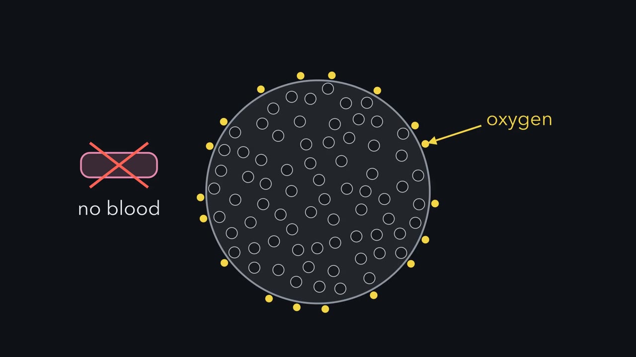
Then came cells from people with Timothy syndrome, caused by a mutation in a calcium channel gene. Patient interneurons made more jumps, but shorter ones. Overall, they moved less efficiently. Fused to control cortex, patient interneurons still showed the defect. So it lay at least partly in the interneurons themselves. Blocking L-type calcium channels improved their migration. Those two experiments, not the channel's location, point to a defect in the cells' own program. An assembloid can show it. A host restores more. Human organoids were transplanted into newborn rat cortex. As the rats matured, the grafts gained a blood supply and responded to whisker deflection.
In 2013, Lancaster and colleagues grew organoids from a person with microcephaly, a brain far smaller than normal. The patient carried mutations in a gene called CDK5RAP2. The patient's organoids had smaller progenitor zones. Is the mutation to blame? Predict. Name three other explanations, and a control that tests each.
The rest of that person's genes. Several donors help, but only moving the gene itself really tests this. A quirk of one cell line. Several clones from the patient test that. A bad batch. Grow patient and control side by side, more than once. These controls strengthen the comparison. Then move the gene itself. Put back into patient tissue, CDK5RAP2 kept more cells as radial glia. Reduced in control tissue by two different hairpin RNAs, it let cells shift toward neurons.
Why was the organoid a good bet? The protein works at the centrosome, inside the dividing progenitor. The rescue and knockdown, done in organoid tissue, made it a result. If a gene acted through blood vessels or microglia, a normal-looking organoid would not clear it. Use the program to choose where to start. Then test the organoid, hardest where the surroundings matter, and measure every feature, because the dish adds stress of its own.
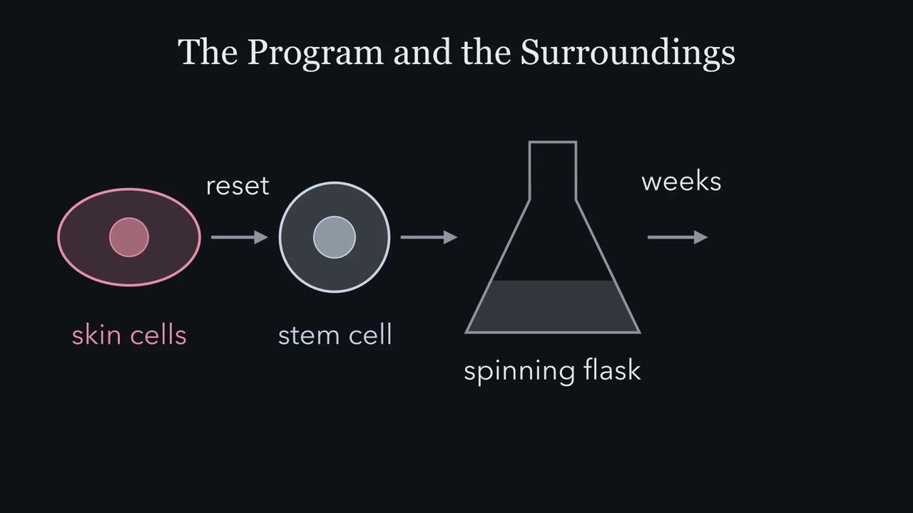
Can a person's skin cells, first reset to stem cells and then grown for weeks in a spinning flask, become a model of their early cortex?
Continue in the lesson: Brains in a Dish: Stem Cells and Organoids →
3:36 · Free, with captions and a transcript. The lesson has the full text, the questions and the sources.
More films from this lesson
How this film was made. Narration: an AI narrator voice (Kokoro, af_heart) reading narration written from this lesson's text. Animation: AI-generated in code (Manim) by Claude, a schematic with no drawn molecular structures. Captions come from the narration script. See something wrong? Report it.