Follow the Markers
Read the transcript of “Follow the Markers”
Film transcript: Follow the Markers
How do you find which compartment holds a protein? Chapter 1 showed how pumps and leaks help compartments keep distinct chemistries. To relate that chemistry to a protein, first find where the protein sits. A fluorescence image can show a protein at the lysosome. But at is not inside. That image alone cannot tell which side of the membrane it is on. So labs also take cells apart. They break cells and separate the pieces by a series of centrifuge spins.
Organelles overlap across these crude fractions; no fraction is assumed pure. To read the fractions, measure markers, proteins whose home is already known. Histone H3 marks the nucleus. Cathepsin D marks the lysosome. Pyruvate kinase marks the cytosol. Each marker spreads across the fractions in its own shape. A protein that stays associated tends to travel with its compartment. So an unknown is assigned provisionally to the marker group whose full profile it most closely shares. In 2016, Georg Borner's group did this with mass spectrometry. They reported localization information for more than 8,700 HeLa proteins.
Curated markers guided organellar assignments. Proteins split between compartments can yield mixed profiles and uncertain assignments. So a match is provisional. You would want a second marker that agrees, repeated fractionations, and an imaging check in intact cells. One more test asks whether an enzyme is enclosed by a membrane. In a 1951 report, de Duve's group found acid phosphatase acting less than expected in fresh fractions. More activity appeared once the particles were damaged. Picture the enzyme sealed in the lumen, its substrate outside and unable to cross.
Now add detergent to open the membranes. What happens to the activity? It rises, as long as detergent does not change the enzyme itself. The substrate can now reach it. Activity hidden behind a membrane is called latency.
Following these hidden enzymes across fractions led his group to propose the lysosome in 1955. De Duve later shared the 1974 Nobel Prize in Physiology or Medicine. The wall that holds a lumen's chemistry also hides what sits inside it. So place the protein first, by its whole profile and independent checks. Then ask what its compartment's chemistry does for it.
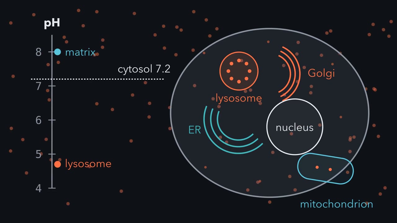
A lysosome near pH 4.7 and a mitochondrial matrix near pH 8 share one cytosol near 7.2. What holds each at its own number?
Continue in the lesson: Organelles as Distinct Environments →
3:20 · Free, with captions and a transcript. The lesson has the full text, the questions and the sources.
More films from this lesson
How this film was made. Narration: an AI narrator voice (Kokoro, af_heart) reading narration written from this lesson's text. Animation: AI-generated in code (Manim) by Claude, a schematic with no drawn molecular structures. Captions come from the narration script. See something wrong? Report it.