Catching Proteins on the Move
Read the transcript of “Catching Proteins on the Move”
Film transcript: Catching Proteins on the Move
How do we know proteins really travel in this order? The first film drew the route, from the rough ER to the Golgi to the cell membrane. But a drawing is not evidence. In 1967, James Jamieson and George Palade found a way to test it. They used thin slices of guinea pig pancreas. The cells they studied make digestive enzymes, not insulin. But they too send proteins out.
Here is the problem. A cell is always making new protein. If every protein carries a mark, the mark is everywhere, and you learn nothing about order. You need to mark just one batch. So they gave the slices a short pulse of a radioactive building block. Only proteins made during those few minutes carried the mark. Then came the chase: plenty of the ordinary building block. Proteins made after that carried no mark. The marked batch was now on its own. They followed the batch in two ways. In one, broken cells were spun in a centrifuge, so their parts settled into layers. In the other, electron microscope pictures showed where radioactive grains sat.
Time for a prediction. The clock reads zero minutes of chase. Right after the pulse, where do you predict most of the marked protein will be? Over the rough ER. At the start of the chase, most grains sat there. That is where ribosomes build new protein. Minutes later, the marked batch had spread out. By seven minutes, Golgi vesicles held almost as many grains as the rough ER. Golgi vesicles are small vesicles at the Golgi's edge. Later still, the mark moved on again. The grains gathered in condensing vacuoles. These are storage granules that are still being formed. At the last time point shown, more than half the grains sat in finished storage granules. These hold the protein until the cell releases it.
Each part's share of the mark peaked in turn. Rough ER, then Golgi vesicles, then condensing vacuoles, then storage granules. The chart shows only four moments of that journey. These were digestive enzyme cells, not beta cells. Still, this experiment gave direct evidence for the order of stops that textbooks still draw. A drawing had become a measurement. So how do we know the order? Mark one batch of new protein, then watch where it goes over time. Each stop's share peaks later than the stop before it.
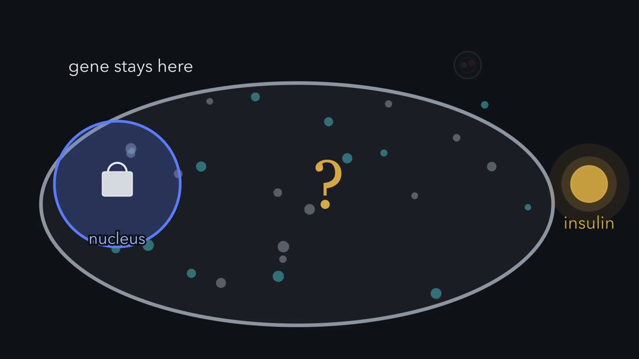
The insulin gene never leaves a beta cell's nucleus, yet insulin leaves the cell after a meal. What carries it out, and in what order?
Continue in the lesson: Organelles as a System →
3:11 · Free, with captions and a transcript. The lesson has the full text, the questions and the sources.
More films from this lesson
How this film was made. Narration: an AI narrator voice (Kokoro, af_heart) reading narration written from this lesson's text. Animation: AI-generated in code (Manim) by Claude, a schematic with no drawn molecular structures. Captions come from the narration script. See something wrong? Report it.