One Tube, Bent Once
Read the transcript of “One Tube, Bent Once”
Film transcript: One Tube, Bent Once
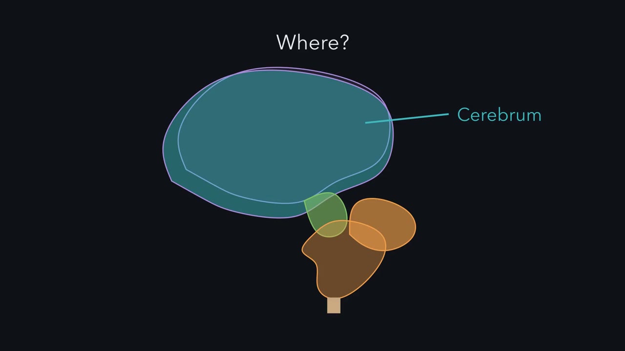
Your brain has a great many named parts. Is one shape behind them all? Most neurons and most glia of your brain and spinal cord come from the wall of one tube. It forms along the embryo's back. The one idea: picture the central nervous system as one hollow tube, bent once. Its walls became the brain's parts and the spinal cord. Its canal became the ventricles. And the tube's own long axis, not the body's, sets what the direction words mean. As it grows, the front end swells into three bulges: forebrain, midbrain and hindbrain. The forebrain and the hindbrain each divide in two. The rest becomes the spinal cord. The front of the forebrain buds out on each side, as the two cerebral hemispheres.
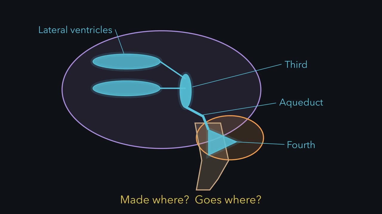
Their canals become the two lateral ventricles. The back of the forebrain becomes the thalamus and hypothalamus, around the third ventricle. The midbrain keeps a narrow canal, the cerebral aqueduct. The hindbrain becomes the pons and medulla, around the fourth ventricle. The cerebellum grows from the hindbrain's dorsal side, so the fourth ventricle lies beneath it. In the spinal cord, many neurons that receive sensory signals develop in the tube's dorsal half, and motor neurons in its ventral half. It is a plan, not a complete split: sensory fibres also reach ventral neurons. So sensory fibres enter through dorsal roots, and motor axons leave through ventral roots.
The brainstem keeps this plan, with sensory nuclei moved to the side. The forebrain does not follow it. In a rat, the tube runs nearly straight from nose to tail. Dorsal is toward its back. Stand the rat up, face pointing forward. The body, cord and brainstem turn. Only the forebrain keeps its frame. In a human, the tube bends by roughly a right angle where the midbrain meets the diencephalon. The embryo bends in three places. This one matters for naming. You are standing. In your spinal cord, which way does dorsal point?
Toward your back. The brainstem uses the same frame. In your forebrain, dorsal points to the top of your head. Three planes name the sections: sagittal, coronal and horizontal. A horizontal cut runs along the forebrain's axis. How does it pass through the brainstem?
Roughly across it, because the brainstem's axis runs up and down. So the tube tells you where things are. What each place does is another question.
Inject a protein tracer into a mouse's blood and, in the cerebral cortex, it goes no farther than the cells lining the capillaries. Why there?
Continue in the lesson: The Map: How the Nervous System Is Organized →
3:05 · Free, with captions and a transcript. The lesson has the full text, the questions and the sources.
More films from this lesson
How this film was made. Narration: an AI narrator voice (Kokoro, af_heart) reading narration written from this lesson's text. Animation: AI-generated in code (Manim) by Claude, a schematic with no drawn molecular structures. Captions come from the narration script. See something wrong? Report it.