What an Overlap Can Claim
Read the transcript of “What an Overlap Can Claim”
Film transcript: What an Overlap Can Claim
Red and green image channels overlap. Does the yellow patch mean the two proteins bind? Chapter one showed why close objects merge into one spot. Now follow the evidence from label to claim. Start with the blur from chapter one.
Across the image, it is about 230 nanometres wide. Along the depth direction, it stretches to about 800 nanometres. A yellow pixel alone cannot show that the two sources are closer than this scale. These dimensions use chapter one's green-light settings. A red channel usually has a longer emission wavelength and a larger blur. For a two-colour claim, use the measured resolution of each channel. Here is a question to hold. At typical cellular protein density, how many protein molecules would this volume contain? A handful, a few hundred, or tens of thousands?
Treat the blur as an ellipsoid. Its volume is roughly 0.02 cubic micrometres. At typical cellular protein density, it contains tens of thousands of protein molecules. So, after the controls, an in-focus overlap can support proximity within about this volume. It does not say the two proteins touch. In widefield, out-of-focus haze can create apparent overlap from farther away. Even that weaker claim needs evidence. Colour overlap is a claim about proximity. It is only as strong as each link that produced it. Check three links in turn.
First, the label. An antibody can bind the wrong protein. Signal loss after knockout supports specificity. With knockdown, the signal should fall with verified target reduction. A secondary-only sample checks nonspecific signal from the fluorescent secondary. Second, the channels. A dye's light can leak into another colour channel. That leak is called bleed-through. Single-label samples measure it, and multicolour beads test whether the channels line up. Misregistration can separate matching signals or superimpose different ones. Third, the record.
Image every condition with identical settings. Keep the relevant pixels below the detector maximum. Background subtraction removes the average background, but not its photon noise. Now return to yellow. If every link holds, an in-focus overlap supports proximity at roughly one resolution volume.
Binding is a separate claim, and it needs other experiments. Co-immunoprecipitation supports membership in the same complex, but not direct contact. A test with purified proteins addresses direct interaction. So read a colour overlap the way chapter one read the blur. Ask what was labelled, what leaked between channels, and what the detector recorded. The overlap reports what the measurement could separate, and nothing finer. Overlap supports proximity, not binding.
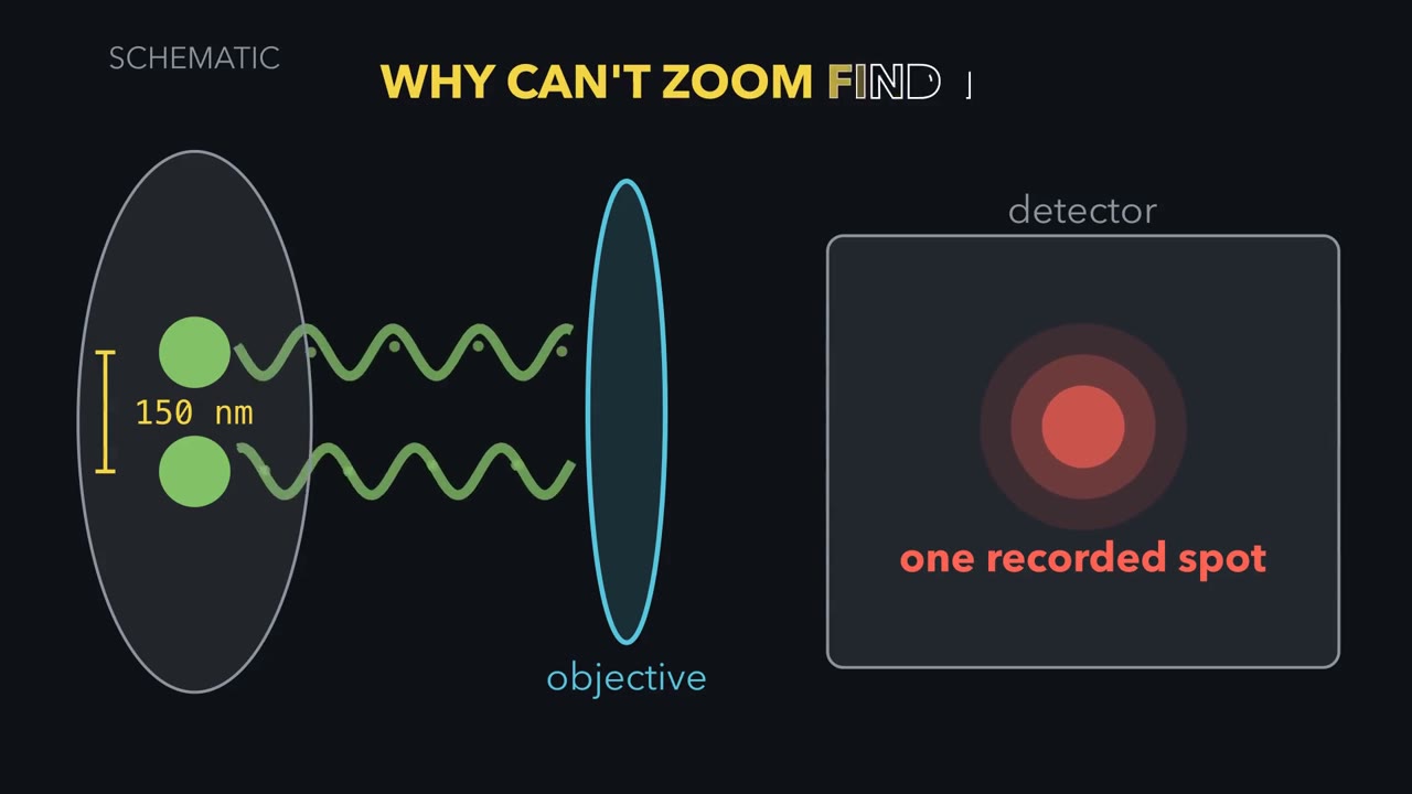
Two GFP-filled vesicles 150 nm apart look like one blob in the best ordinary light microscope. Zooming in will not split them. Why not?
Continue in the lesson: Microscopy and Quantitative Images →
3:42 · Free, with captions and a transcript. The lesson has the full text, the questions and the sources.
More films from this lesson
How this film was made. Narration: an AI narrator voice (Kokoro, af_heart) reading narration written from this lesson's text. Animation: AI-generated in code (Manim) by Claude, a schematic with no drawn molecular structures. Captions come from the narration script. See something wrong? Report it.