What Zoom Cannot Recover
Read the transcript of “What Zoom Cannot Recover”
Film transcript: What Zoom Cannot Recover
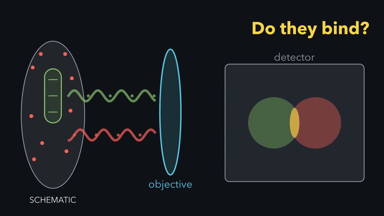
These two GFP-filled vesicles are 150 nanometres apart. An ordinary light microscope records one spot. Why can software zoom not split it back into two? Follow the light from specimen to screen, and watch where the missing detail is lost.
Start with one point source. In ideal widefield fluorescence, every point becomes a blur. The screen shows a bright centre with faint rings, an Airy pattern. This computed green curve is its intensity across the screen. Why does the point spread?
Light diffracts at the objective opening. A finer pattern bends light through wider angles. The lens collects only a cone, so some information from fine detail misses the objective. Numerical aperture describes the width of the collected cone. In this ideal lateral model, the Rayleigh distance equals 0.61 times emission wavelength, divided by numerical aperture. Emission wavelength is the colour released by the fluorescent label. In the ideal lateral model, the Rayleigh distance gets smaller with shorter emission wavelength or higher numerical aperture. It is one resolution convention, not a universal border. Now restore both vesicles. Their separation is 150 nanometres.
For the settings shown here, the displayed Rayleigh distance is about 227 nanometres. Will the detector show two peaks, or one? Each source still makes its own blur. On the detector, their intensities add point by point. The two computed curves merge into one summed peak. At one hundred fifty nanometres, no dip survives between them. They are unresolved here, although other methods may separate them. Move the sources apart. A shallow dip opens before they reach the Rayleigh distance. At that distance, the central dip is about one quarter of peak height. That clear dip is the convention for calling two points resolved. For two ideal points, the Rayleigh distance marks a clear dip, not the first appearance of two peaks.
Return the vesicles to 150 nanometres. The detector again records one peak. Software zoom enlarges that record. It cannot add detail the optics and camera never captured. Optical magnification before the camera can change sampling; that is a different operation. Bigger display pixels cannot recover a missing dip. To separate or interpret closer objects, change how information enters the record. A confocal pinhole asks which out-of-focus light reaches the detector. Super-resolution asks how illumination or glowing molecules can encode closer detail. Electron microscopy replaces light with a different probe. Each change answers a measurement problem and brings new limits.
A microscope image is a measurement, not the specimen itself. Before trusting one, ask what was labelled, what the instrument could separate, and what the detector recorded. Software zoom makes the same record bigger. Software zoom cannot add detail the optics and camera never captured.
Two GFP-filled vesicles 150 nm apart look like one blob in the best ordinary light microscope. Zooming in will not split them. Why not?
Continue in the lesson: Microscopy and Quantitative Images →
3:55 · Free, with captions and a transcript. The lesson has the full text, the questions and the sources.
More films from this lesson
How this film was made. Narration: written from this lesson's text and read by an AI narrator voice (Kokoro, af_heart). Animation: AI-generated in code (Manim) by Claude, a schematic with no drawn molecular structures. Captions come from the narration script. See something wrong? Report it.