Counting Dots to Measure a Cell
Read the transcript of “Counting Dots to Measure a Cell”
Film transcript: Counting Dots to Measure a Cell
Every job needs the team. But how much of a cell is each part? You can't pour a cell into a measuring cup. So how do scientists find out how much of a cell is mitochondria? First, they preserve the cell and cut a very thin slice. A transmission electron microscope sends a beam of electrons through the slice. It shows small organelles that a school microscope cannot. Because the cell was preserved, each picture is a still view, not a living cell. Next comes a simple trick called point counting. Lay an even grid of dots over the picture. Then look at where each dot lands. Dots that land on mitochondria turn gold. One slice gives only a rough guess.
Over many fairly chosen pictures, the share of gold dots estimates the share of the cell's volume that mitochondria fill. Try it on a small grid. Count the gold dots, then count all the dots. The share appears on the screen. This toy grid lands inside the real range for heart muscle. Real studies used many pictures and many more dots. Look at heart muscle cells from ten kinds of mammals.
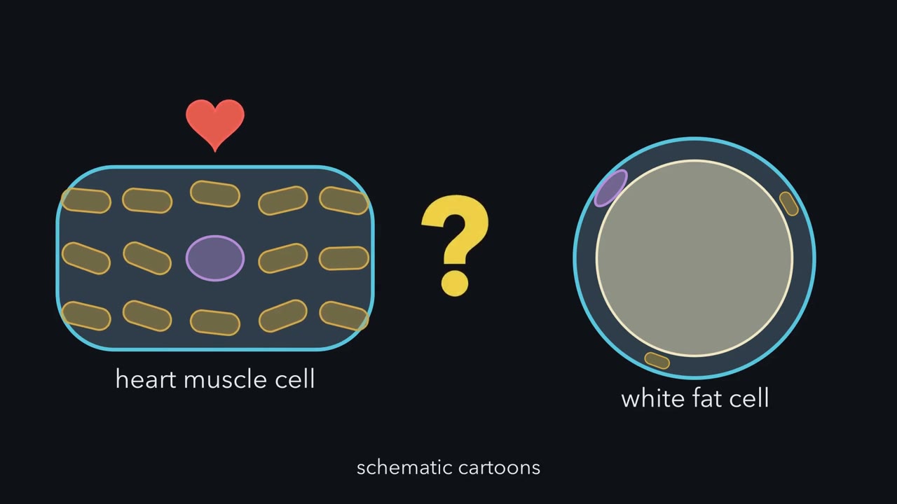
Mitochondria filled about a fifth to over a third of the cell, depending on the kind of mammal. Now drop the same grid on a white fat cell. Before the dots turn gold, predict. Will its share be bigger or smaller than the heart cell's?
Much smaller. Almost every dot lands on fat. One team laid a grid of 682 points over pictures of white fat cells from chicken embryos. Fat droplets filled about nine tenths of the cell. Mitochondria filled less than one percent. The fat takes most of the floor. Everything else, mitochondria included, sits in a thin rim around it. Is that rim short of mitochondria? The lesson lets you work it out. So in the heart cell, mitochondria fill a large share. In the fat cell, stored fat fills most of the space. Many of the same parts, different floor plans. One caution. These cells came from different animals and different studies. They show a pattern across cells with different jobs.
They do not prove what causes it. Still, simple counting, repeated over many pictures, turned images into numbers. Those numbers show a pattern: cells with different jobs have different floor plans.
Why is a heart muscle cell crowded with mitochondria, while a fat cell is mostly one big drop of fat?
Continue in the lesson: Inside a Cell →
3:06 · Free, with captions and a transcript. The lesson has the full text, the questions and the sources.
More films from this lesson
How this film was made. Narration: an AI narrator voice (Kokoro, af_heart) reading narration written from this lesson's text. Animation: AI-generated in code (Manim) by Claude, a schematic with no drawn molecular structures. Captions come from the narration script. See something wrong? Report it.