How a Receptor Loads a Switch
Read the transcript of “How a Receptor Loads a Switch”
Film transcript: How a Receptor Loads a Switch
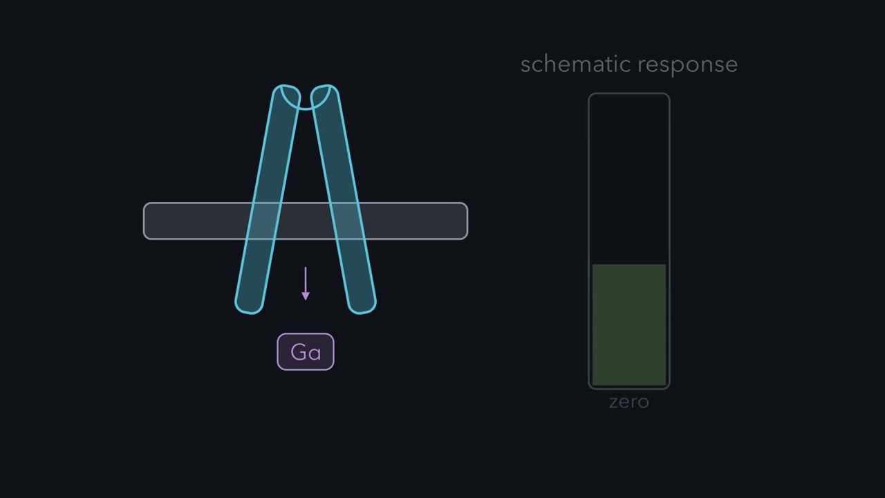
How can binding outside a cell load a molecular switch inside it? Follow one membrane as the message crosses without the ligand crossing. A G-protein-coupled receptor, or GPCR, is one chain woven repeatedly through the membrane. Its outer pocket and inner face are physically connected. An agonist is a ligand that favors receptor activation. It settles into the pocket and favors an active shape. One cytoplasmic helix swings outward, opening a cavity below. A membrane-bound G protein waits below as a trimer. Purple Gα alpha holds gray GDP.
Teal βγ stays pressed against it. In this resting arrangement, the switch is off. The Gα alpha tail rises into the receptor cavity, bringing the whole trimer to the active receptor. Docking changes Gα's grip on GDP. The gray nucleotide leaves first. Green GTP can then occupy the empty site. The receptor has acted as an exchange factor, not as a source of GTP. It remains ready to load another G protein while agonist stays bound. GTP changes the Gα -alpha surface and frees βγ to engage targets. The textbook picture draws them apart.
Activated Gi may instead rearrange without fully separating. Either way, both surfaces can regulate effectors. Hydrolysis later returns Gα -alpha to GDP and favors reassembly. Now the cell chooses what those surfaces meet.
Follow the hourglass. Many receptors narrow to a few Gα -alpha families and βγ, then widen toward enzymes and channels. One branch raises cAMP. Another lowers it. Another raises calcium. βγ can open a potassium channel. Each family points to a main direct target, not a guaranteed exclusive partner. The receptor makes the first selection. The cell then supplies whichever enzymes, channels, and scaffolds are present. So one messenger can carry different meanings in different cells. The same acetylcholine can therefore meet different wiring. In a pacemaker cell, the receptor on screen loads Gi, and freed βγ opens potassium channels. Firing slows.
In airway muscle, a different receptor loads Gq, and phospholipase C-β -beta raises calcium. The muscle contracts. The input matches; the cellular paths differ. So do not predict an answer from the ligand alone. Trace the receptor subtype, the G protein it loads, and the targets available. That path predicts the cell's first response.
Dose and efficacy still change how strongly the receptor is activated. The wiring decides what that activation means.
Adrenaline strengthens the heartbeat but relaxes the airways. How can the same hormone give opposite orders?
Continue in the lesson: GPCRs and Trimeric G Proteins →
3:36 · Free, with captions and a transcript. The lesson has the full text, the questions and the sources.
More films from this lesson
How this film was made. Narration: written from this lesson's text and read by an AI narrator voice (Kokoro, af_heart). Animation: AI-generated in code (Manim) by Claude, a schematic with no drawn molecular structures. Captions come from the narration script. See something wrong? Report it.