The Seal and the Sides
Read the transcript of “The Seal and the Sides”
Film transcript: The Seal and the Sides
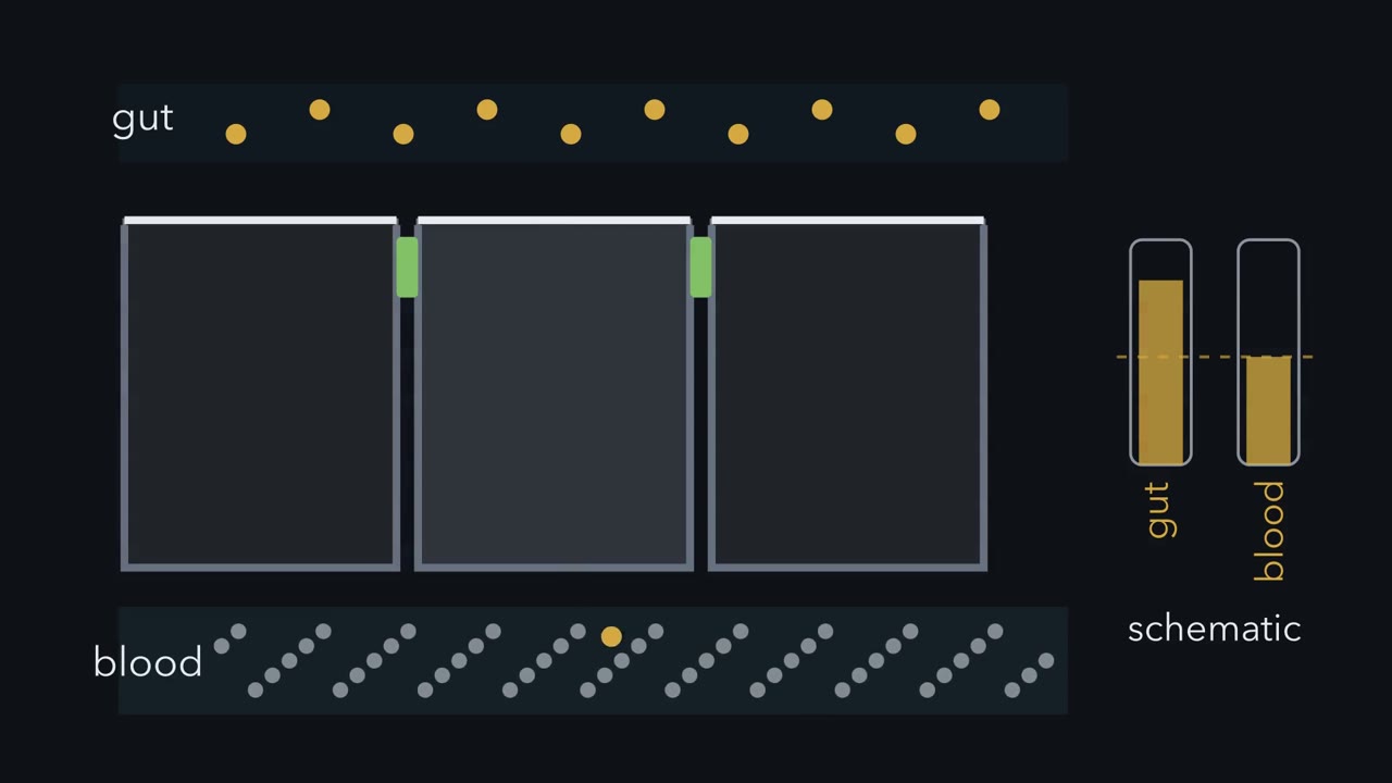
What makes the seal between cells selective, and what keeps each cell's two faces distinct? In chapter one, under gut gradients, carrier placement biased glucose toward blood. The seal decided what crossed between cells.
The seal is the tight junction, a belt where neighbouring cells meet near their apical edges. It is not solid caulk. It is a selective gate between cells. Claudin proteins assemble into branching strands around each cell. Narrow routes through those strands can pass small ions.
Meanwhile, the junction strongly restricts bacteria and larger molecules. The small intestine needs some sodium and water to cross between cells, so its junctions are relatively leaky. The bladder needs a tighter barrier. Different epithelia tune this gate for different jobs. How can we measure this gate? Grow cells as a sheet on a porous filter. Put one electrode above and another below, then pass current. If resistance falls, must glucose be leaking through?
No. This reading is transepithelial electrical resistance, or TEER. It reports how easily ions cross the whole sheet. It cannot name the ion or route, or say whether larger molecules leak. Current may cross between cells or pass through them. A low reading can reflect selective ion pores, broader leaks, or breaks. Telling them apart takes other experiments, set as a design problem below. The seal is often called a fence, said to stop membrane proteins drifting between the two faces. How well it fences is tested in the text below.
Cells also mark territories with proteins, and traffic delivers cargo to each face. In the standard scheme, Crumbs marks the apical side, while Scribble marks the basolateral side. The Par complex sits near their boundary. These groups are thought to restrain one another, while polarized traffic places carriers. Below the seal, an adherens junction couples neighbouring actin belts. Desmosomes link keratin networks against stretching.
Gap junctions pass ions and small molecules between cytosols. At the base, hemidesmosomes connect keratin to the basement membrane. Each junction has a different job. Together they let a one-cell-thick sheet stay selective, strong, connected, and anchored. Back in the gut, SGLT1 stays apical while the pump stays basolateral. The selective seal controls the path between cells, while deeper junctions keep the sheet intact. That is the rule: epithelial barriers are built by division of labour. A gate, distinct faces, grips, anchors, and channels work together, but they are not interchangeable.
The gut lining is one cell thick. How does it keep pulling glucose into the blood, even when the gut holds less than the blood does?
Continue in the lesson: Epithelia and Tissue Organization →
3:27 · Free, with captions and a transcript. The lesson has the full text, the questions and the sources.
More films from this lesson
How this film was made. Narration: an AI narrator voice (Kokoro, af_heart) reading narration written from this lesson's text. Animation: AI-generated in code (Manim) by Claude, a schematic with no drawn molecular structures. Captions come from the narration script. See something wrong? Report it.