The Band Before the Furrow
Read the transcript of “The Band Before the Furrow”
Film transcript: The Band Before the Furrow
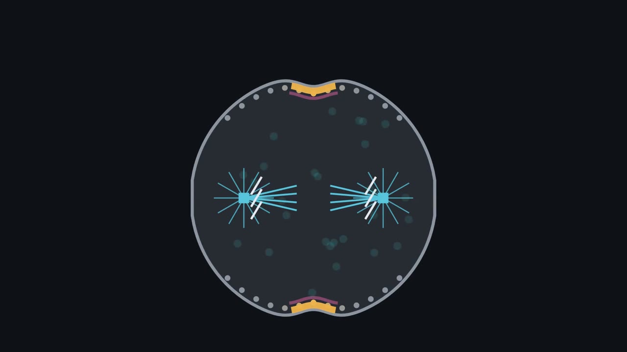
This sea urchin embryo cell is in anaphase: its two sets of chromosomes are moving apart. Now it must cut itself in two, right between them. How does its surface know where the middle is? The cut happens at the cell surface. There the surface pinches in, making a groove called the furrow. But the thing that marks the middle sits deep inside. It is the spindle, a bundle of microtubules with a star-shaped aster at each end. There are three simple answers. One: the spindle pulls the surface in directly, by force. Two: an actin ring gathers at the middle on its own. Three: the spindle only sends a signal. The signal marks the surface, and the ring is built under that mark.
To tell them apart, researchers watched a switch protein called RhoA. RhoA is on when it holds GTP, and off when it holds GDP. They added a glowing probe that sticks only to switched-on RhoA. A band of active RhoA, shown here in amber, lights up just under the membrane. One to four minutes later, the furrow starts underneath it. The same order was seen in frog embryos. The mark comes before the cut. Next, they blocked actin assembly with a drug, so no ring could be built. In sea urchin embryos, the amber band still formed. So the signal does not need the ring. Answer two is out. Now the decisive test. This cell has already begun to furrow. A blunt needle pushes the spindle to one side.
What moves first: the furrow, the amber band, or nothing? Over a few minutes, active RhoA gathers over the spindle's new middle. Only then does the furrow follow it there. The band leads, and the furrow follows. That order argues against the spindle simply dragging the surface, and it supports answer three: signal first. Here is the rule for these embryo cells. The cut goes where the anaphase microtubules place a band of active RhoA. Move the microtubules, and the band moves first. The furrow follows. Under the band, active RhoA turns on formins and promotes myosin II. Formins build long actin filaments.
Myosin II, a motor, pulls those filaments past each other. In its simplest picture, the result is a ring that tightens like a drawstring. Now the two daughters are joined only by a thin bridge. Cutting that bridge is a separate job. One question stays open: how do microtubules deep inside switch on a protein at the membrane? But where the cut went was settled earlier, where the microtubules placed the band.
Push the spindle off-centre in a sea urchin embryo cell, and the signal for the furrow follows it before the furrow does.
Continue in the lesson: Cytokinesis and Organelle Inheritance →
3:26 · Free, with captions and a transcript. The lesson has the full text, the questions and the sources.
More films from this lesson
How this film was made. Narration: written from this lesson's text and read by an AI narrator voice (Kokoro, af_heart). Animation: AI-generated in code (Manim) by Claude, a schematic with no drawn molecular structures. Captions come from the narration script. See something wrong? Report it.