Ruling Out the Dull Explanations
Read the transcript of “Ruling Out the Dull Explanations”
Film transcript: Ruling Out the Dull Explanations
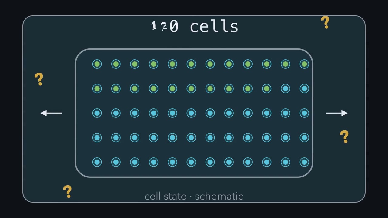
Stretched dishes show more tracked divisions. What else, besides stretch, changed in those dishes? Chapter one locked three things: the prediction, endpoint and independent unit. A control is how you find out. Look at what the stretched dish went through. Someone assembled the device, mounted it and carried it to the microscope. If the control dish skipped those steps, stretch now includes every handling difference. So unstretched devices are assembled, mounted and imaged identically. The medium raises the same problem. Wnt3A medium comes from mouse L cells engineered to make Wnt3A. The matched control is medium from the parental L cells, prepared the same way.
Even that cannot promise Wnt3A is the only difference. So add a readout of the pathway itself. TOPdGFP is a short-lived green reporter driven by beta-catenin-responsive sites. Its signal asks whether the Wnt medium engaged beta-catenin-dependent transcription in these cells. If it stays flat, the medium may not have done its job. Each of these follows one rule. A good control matches the treatment in every step except the one being tested. Without that match, the dull explanation and the interesting one predict the same result. Then a difference between them can point to that step. A drug control gets the vehicle, the drug's solvent without drug, at the same concentration. Whoever scores the cells fixes positivity thresholds before analysis and scores blind to condition.
Otherwise expectations can slip into the counts, one borderline cell at a time. Stretch itself must be checked. Measure the strain the cells actually feel, and analyze only the strained region. In the 2016 study, two biaxial devices reached about 8.5% and 15%. percent. Those results should not be merged. Each device is its own condition. Now predict. Suppose stretched cells stall, and you suspect DNA damage. You stain for a damage marker, and it stays blank. Does that blank marker show there was no damage?
Only if the assay could have seen damage. Benham-Pyle and colleagues ran methyl methanesulfonate, a DNA-damaging chemical, as a positive control. It shows the assay detects damage when damage is present. Even then, one missing marker cannot rule out every kind of damage. Return to the stretched dish and its unstretched twin. They share the handling, medium, vehicle and blinded scorer. The strain itself was measured. Only the stretch differs. Now a difference in tracked divisions can point to stretch, for the period watched.
Stretched cells glow green. What would it take to show that stretch made them divide?
Continue in the lesson: Designing a Cell Biology Experiment →
3:30 · Free, with captions and a transcript. The lesson has the full text, the questions and the sources.
More films from this lesson
How this film was made. Narration: an AI narrator voice (Kokoro, af_heart) reading narration written from this lesson's text. Animation: AI-generated in code (Manim) by Claude, a schematic with no drawn molecular structures. Captions come from the narration script. See something wrong? Report it.