Biology · Skim version
How the DNA Model Was Built
Read the full lesson → Skim the whole course →
The guiding idea
Four pictures can all represent the same DNA. Which one should you trust?
Choose DNA models by the question; every model leaves features out.
From the film: Which Picture of DNA Should You Trust?
- Choose DNA models by the question; every model leaves features out.
- A ladder shows base pairing, while a letter string shows base order.
- Base amounts, X-ray measurements and chemistry constrained the double-helix model.
- A diffraction pattern gives indirect measurements, not a photograph of DNA.
- Watson and Crick called the model unproved; later tests supported its main features.
From the film: How DNA Became Readable
- Sequencing reads base order, which a shape model does not show.
- Stopped copies sorted by length, shortest first, reveal a complementary base sequence.
- A reference sequence is a comparison standard, not one person's genome.
- Lower production costs let biologists compare many genomes.
-
Explore: four models of one molecule
-
What each model can answer
-
Three kinds of evidence, one model
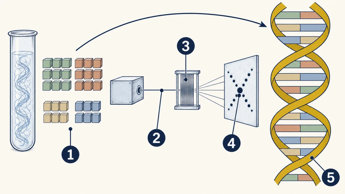
- Base amounts
- X-rays
- lined-up DNA fibers
- pattern of spots
- double helix
-
Reading the letters since 1977
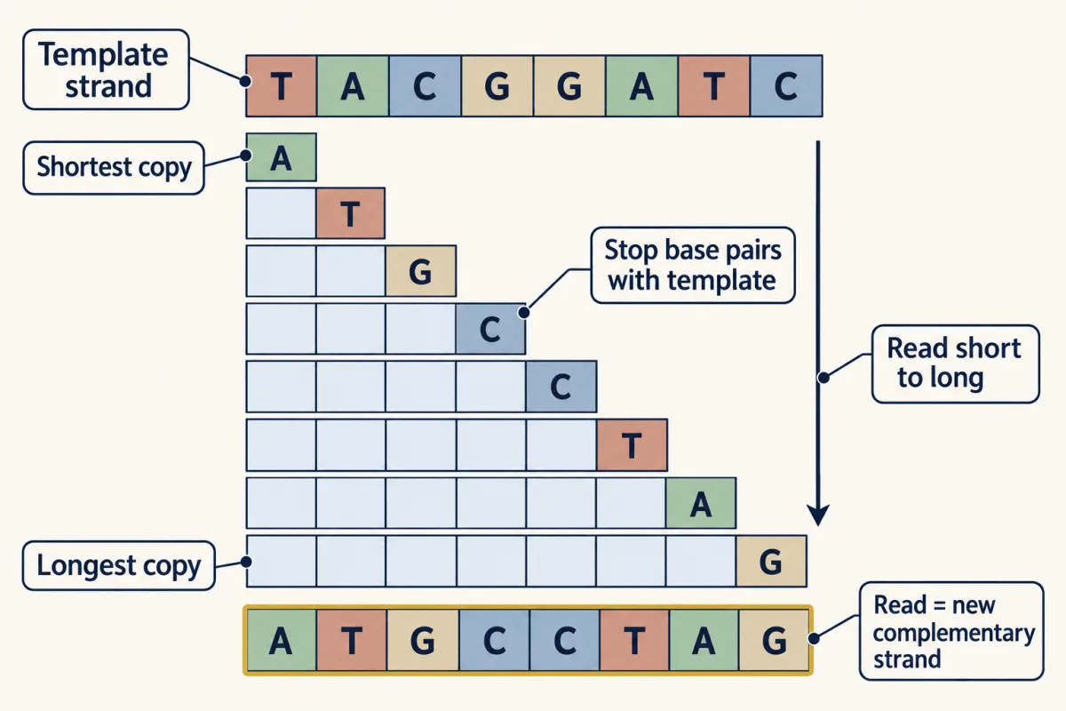
The idea behind Sanger's 1977 method: copies of a strand stop at each base, and their lengths reveal the order of the bases. AI-generated illustration -
The falling cost of a genome
-
Evaluate: check your thinking
Where this lesson fits
Before thisReview first
- DNA: Structure and CopyingThis course · Lesson 16
You need base pairing and the two-strand structure from there to judge what each DNA model shows.
- What Am I Made Of?This course · Lesson 2
You need to know nucleic acids are chains of nucleotide building blocks, which this lesson's models draw.
After thisStudy next
- From DNA to ProteinThis course · Lesson 18
The base order that sequencing reads is the code that gets copied and translated into protein here.
- Engineering GenesThis course · Lesson 22
Reading and cutting DNA sequences is what lets scientists move a gene into bacteria here.
Suggested by an AI model from the lessons themselves, then checked. How we use AI