How to Prove a Gut Synapse
Read the transcript of “How to Prove a Gut Synapse”
Film transcript: How to Prove a Gut Synapse
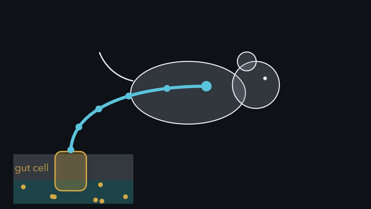
A cell in the gut lining is not a neuron. How could it send a rapid message to a vagal sensory neuron? One possibility is a hormone released into nearby tissue. Another is a point-to-point synapse. Speed alone will not settle the choice. Start with anatomy.
Modified rabies virus begins in selected enteroendocrine cells and labels connected nerves. In some mice, labelling reaches vagal nodose neurons. Tracing can map a connection across the epithelial boundary. It cannot show rapid transmission, identify the messenger, or reveal whether the connection changes nerve firing. The picture must become physiology. That establishes connectivity, not the transmitter. The cells also carry transcripts for VGLUT1. Those transcripts fit glutamate release, but still do not show a current. Now move the cell and a nodose neuron into a dish. Light activates the gut cell. Within 60 to 800 milliseconds, an excitatory current appears in the neuron.
That current measures the neuron's response directly. Its sub-second latency supports a fast messenger in this dish. But one current still cannot establish which receptors carry it. Kynurenic acid blocks ionotropic glutamate receptors, and the current disappears. If receptors are blocked rather than cells damaged, what should happen after washout?
Washout removes the blocker, and the current returns. Recovery supports receptor block rather than cell damage. Together, latency and reversible pharmacology identify fast glutamate action on the neuron. Here is the rule. No single result proves the route. A fast synapse is established when anatomy, timing, pharmacology and causal perturbation converge. Causality comes from the living mouse. Intestinal sucrose raises cervical-vagus firing. Blocking SGLT1 removes that response, placing the sugar sensor upstream. Light that silences CCK-labelled gut cells abolishes the sucrose response.
Light that activates the same labelled population raises vagal firing. These tests ask necessity and sufficiency. The chain has boundaries. Cocultures are dishes. Only a minority of CCK cells contacted sensory fibres in fixed tissue. A synapse does not erase hormone signalling. Anatomy found the contact. Physiology identified rapid glutamate transmission. Acute manipulations connected the sensor to vagal firing. The next question is what this signal changes.
A mouse with no sweet taste still learns to prefer sugar over a sweetener. The sensor is in its gut, and it is not a neuron. How would you show that?
Continue in the lesson: Sensing the Gut: Enteroendocrine Cells and Nutrient Signals →
3:29 · Free, with captions and a transcript. The lesson has the full text, the questions and the sources.
More films from this lesson
How this film was made. Narration: written from this lesson's text and read by an AI narrator voice (Kokoro, af_heart). Animation: AI-generated in code (Manim) by Claude, a schematic with no drawn molecular structures. Captions come from the narration script. See something wrong? Report it.